Testicular cancer is very common in intact male dogs
Most tumors are contained to the testicles, but may spread in 10 to 20% of the cases
Surgery is the the treatment of choice
Prognosis is usually very good
Introduction
In one study showed that 27% of male dogs develop testicular tumors.1
Sertoli cell tumors, interstitial (Leydig) cell tumors, and seminomas are the three most common tumors. Seminomas (42%) and interstitial cell tumors (50%) are most common with sertoli cell tumors being the least common (8%). About one third of dogs that develop a tumor will have more that one of these types of tumors present. Other types of testicular tumors (i.e. embryonal carcinoma, lipoma, fibroma, hemangioma, chondroma, teratoma) can occur, but are rare.
The current cause of testicular tumor development is unknown. Male dogs that have one or both testicles that have not descended from the belly cavity are much more likely to develop a tumor than dogs with normal (scrotal) testicles.
Undescended testicles (located in the abdomen or groin) are at increased risk to develop malignant behaving tumors (seminomas and Sertoli cell tumors). Tumors of normal descended, or scrotal, testicles are usually benign. The tumor is slow to metastasize and the common site of spreading include lymph nodes.
Testicular tumors can spread to regional lymph nodes, liver and lungs.
Specific tumors characteristics
Sertoli cell tumors
higher rate of spread than other testicular tumors
more common in undescended testes.
Interstitial (Leydig) cell tumors
are benign and small
show very few symptoms
these tumors are usually incidental findings
Seminomas
arise from the cells of the testicle that normally produce sperm
majority are benign and they rarely spread.
may produce estrogen and result in feminization
Clinical Signs
Any breed can be affected and are found in older dogs (greater than 10 years) and obviously in intact males
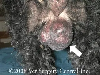
Swelling of one or both testicles
Generalized scrotal enlargement
Infertility in the breeding stud
Effects of excessive estrogen levels due to tumor:
Symmetrical hair loss
Brittle hair
Poor hair regrowth
Thin skin
Hyperpigmentation (darkening of the skin)
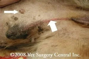
Stripe of red inflammation along the midline of the prepuce (large arrow in photo right)
Nipple elongation (small arrow in photo right)
Mammary enlargement
Penile atrophy
Preputial swelling and sagging
Testicular atrophy of the unaffected (noncancerous testicle)
Prostatic atrophy or enlargement
Anemia
Behavioral changes:
Nipple
Squatting to urinate
Reduced sex drive
Attraction of other male dogs
Diagnosis
A thorough physical examination including palpation of the testicles for a mass; the stripe of inflammation seen along the prepuce (arrow in the photo above) is classic for an estrogen producing tumor
Complete blood count (CBC) – to check for anemia
Biochemistry profile – to check internal organ health prior to surgery
Urinalysis +/- culture and sensitivity – to check for concurrent bladder infection
Chest and abdominal radiographs (x-rays) – to check for spread of tumor
Abdominal and scrotal ultrasound – to check for tumor and spread of tumor
Fine needle aspiration or biopsy – not commonly performed, but may be helpful to arrive at a diagnosis prior to surgery
Biopsy of the testicle after it is surgically removed
Treatment
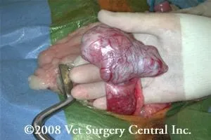
Surgical removal of the testicles is performed. This surgery is typcially very routine, however the scrotal skin should also be removed to prevent the development of a large painful swelling of the scrotum after surgery.
Chemotherapy can be pursued if the tumor has metastasized. Treatment of metastatic disease should be pursued.
Radiotherapy
Potential Complications
The most common complication is marked swelling of the scrotum if it is not ablated at the time of castration.
Infection is uncommon
High estrogen levels may result in bleeding and anemia due to bone marrow suppression
Spread of the tumor will ultimately cause death of the patient
Care after surgery
Check the incision daily for 14 days for signs of infection: swelling, redness, pain or discharge.
A recheck in 10-14 days following surgery is recommended to evaluate incision healing.
If bone marrow disease is present follow-up blood work will need to be performed to monitor for improvement in red blood cell, white blood cell, and platelet numbers.
Patients with malignant tumors should be reevaluated every three to four months for recurrence or metastasis.
Prognosis
Surgery is curative for most testicular tumors. About 10 to 20% of the cases have spread at the time of diagnosis.
Interstitial cell tumors and Sertoli cell tumors without spread or damage to the cells of the bone marrow have an excellent prognosis.
Seminomas without signs of hyperestrogenism also have an excellent prognosis.
Damage to the cells of the bone marrow (caused by the excessive estrogen levels) can be fatal despite therapy, but usually improves two to three weeks after tumor removal.
The prognosis for testicular tumors that have spread is more guarded, but varies greatly depending on the location, type of tumor, and treatment options.
References
Grieco V, Riccardi E, Greppi GF, et al. Canine testicular tumours a study on 232 dogs. J. Comp Path 138: 86-89, 2008.
McDonald RK, Walker M, Legendre AM, et al. Radiotherapy of metastatic seminoma in the dog. J Vet internal Med 2:103-107, 1988.
Dhaliwal RS, Kitchel BE, Knight BL, et al. Treatment of aggressive testicular tumors in four dogs. J Am Anim Hosp Assoc 35: 311-318; 1999.