

Key Points
HOD is a condition that affects young large breed dogs
It is a very painful condition
Medical therapy is required to treat this condition
Prognosis is fair to poor due to recurring episodes of the condition
Dwarfism is a common sequel following moderate to severe HOD
Anatomy
The ends of bones of growing puppies have a zone called the growth plate. The bone adjacent to the growth plate is soft and very developmentally active.
Cause of hypertrophic osteodystrophy
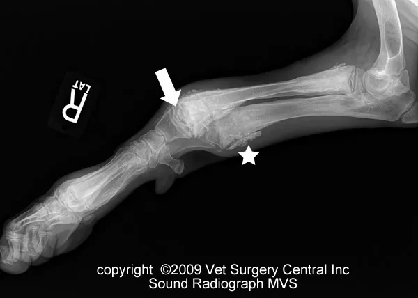
The cause of hypertrophic osteodystrophy in dogs is largely unknown. Proposed causes include distemper virus infection, vaccination with distemper virus, bacterial infection and other viral infections. Vitamin C deficiency is unlikely to be a cause of this disease, as previously believed. A series of events take place at the microscopic level within the affected bones. First, the blood vessels near the growth plate become distended and bleed into the bone. Next, the bone in this region dies, gets resorbed and develops microfracturing due to weakening of the bone structure (photo: see arrow). In response to this, new bone is laid on the surface of the bone (photo: see star).
Signs and diagnosis
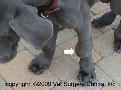
Clinical signs of hypertrophic osteodystrophy include fever, anorexia, and depression. Lameness is always seen with this disease and may vary from mild to severe. With multiple limbs affected, the patient may be reluctant to stand or walk. Typically, the ends of the long bones become thickened (see photo below left). The radius, ulna and tibia bones are most commonly affected; however, the ribs, jaw bone and the bones in the paw can also be affected. Other clinical signs may include diarrhea, discharge from the eyes, tonsillitis, thickening of the footpads, pneumonia and abnormal development of the enamel of the teeth. This condition is commonly seen in rapidly growing large and giant breed dogs from 2 to 6 months of age. Common breeds affected include Great Danes, Boxers, German Shepherds and Weimaraners.
The diagnosis of HOD is based on the presence of supporting clinical signs and findings on x-rays. These x-ray signs include a line of lucency where destruction of the bone adjacent to the growth plates. Sometimes new bone production is also seen on the outside of the bones. Occasionally the growth plate adjacent to the HOD lesion will be damaged which subsequently will result in a bowing of the bone.
Treatment
Treatment is only supportive for hypertrophic osteodystrophy. When a patient goes through an acute phase of the disease and fever is present, intravenous fluids are usually required to keep the patient hydrated. Nutritional support is provided with a feeding tube if the patient refuses to eat for 5 or more days. Pain is controlled with narcotics and nonsteroidal anti-inflammatories. Antibiotics are indicated if the patient has signs of pneumonia or other bacterial infections. If the bones become twisted due to growth plate damage, corrective surgery may be indicated. Because the distemper vaccination has been implicated, inoculation should be delayed until the pet has been in remission for a couple of months. Home care At home, prescribed medication should be given to minimize pain. Exercise should be limited to short leash walks on soft surfaces such as grass. To keep the muscles toned, swimming or exercise in an underwater treadmill may be recommended under the direction of a professional therapist. Monthly evaluations should be made by a veterinarian to check your companion’s health. During each visit, x-rays of the limbs are used to monitor the healing process of the affected bones until the patient is 8 to 10 months of age.
Results
Hypertrophic osteodystrophy is a self-limiting disease that can last for a few weeks per episode. Recurrence of the condition is expected in most affected dogs until the pet is 8 to 10 months of age. The moderate to severe form of this disease will cause dwarfism. Patients that are severely affected by the disease and are unresponsive to treatment should be euthanized.
